
دراسة الدم ومكوناته وخصائصه الفيزيائية, الكيميائية, التشريحية (لأنه يعتبر نسيج حي) والفيسيولوجية تعتبر من المواد المهمة جدا, نظرا لما يقوم به الدم من وظائف حيوية أساسية في جسم الانسان حيث أنه النهر الحي في جسمنا, ناقلا معه ما تيسر من مواد وتركيبات ووظائف أعجز شخصيا أنا أو غيري عن وصف أهميتها ومدى تعقيدها بشكل حصري وكامل ويحتاج الى مجلدات فعلا للتعمق في أموره وخصائصه, وتأتي دراسة وفهم فيسيولوجية وتشريح الدم صيدلانيا لأهميته القصوى في حركية الدواء في جسم الانسان ومدى تأثره سلبا وايجابا بوضع الدم بشكل عام, كما ان دراسته (بالاضافة الى باقي أجزاء الجسم) بشكل مفصل يعيننا على فهم أكثر تعمقا للطب والصيدلة بوجه عام
في هذا القسم الآن سنتحدث بشكل أكثر تخصصا ومتابعة للمقالات السابقة حول طريقة ولادة وتكون كريات الدم الحمراء من نخاع العظم, وآلية التكوين وخطواته المتتالية وأهمية ذلك وكيفية تأثيره على ظهور بعض الأمراض في بعض الأحيان
في هذا القسم الآن سنتحدث بشكل أكثر تخصصا ومتابعة للمقالات السابقة حول طريقة ولادة وتكون كريات الدم الحمراء من نخاع العظم, وآلية التكوين وخطواته المتتالية وأهمية ذلك وكيفية تأثيره على ظهور بعض الأمراض في بعض الأحيان
دراسة ومراجعة اسماعيل العبد مرتضى
يمكن تزويدكم بالمراجع المستخدمة تحت الطلب ويرجى الضغط على الصور لتكبيرها لقراءة محتوياتها

In last lectures, we spoke about the blood in general & about the RBC’s, which are the Red Blood Cells, in this part we’re going to go more deeper in discussing the production process of the RBC’s which is called "Hematopoiesis", or also called "Hemopoiesis"; and this is the major process to get a new born or new made red blood cells
The Bone Marrow

The bone marrow briefly is the location were RBC’s are made; it is composed basically of a large soft network of Reticular Connective Tissue that borders on wide blood capillaries called "Blood Sinusoids". Within this network there are some of the following elements
A) Immature blood cells
B) Macrophages
C) Fat cells
D) Reticular cells; which are the fibroblasts that secrets the fibers
B) Macrophages
C) Fat cells
D) Reticular cells; which are the fibroblasts that secrets the fibers
As it is defined normally that there are 2 type of bone marrows; one is the Red Bone Marrow & the other is the Yellow Bone Marrow, both have similar characteristics in which the red one is consisting mainly of Myeloid Tissues & the yellow one is consisting mainly of fat cells; but now we are concerned more about the Red Bone Marrow as it is the place were the RBC’S, WBC’S & platelets are formed & manufactured, In adults, Red Bone Marrow is found chiefly in the bones of the axial skeleton & gridles, & in the proximal epiphyses of the humerus & femur
Each type of blood cell is produced in different numbers in response to changing body needs & different regulatory factors. As they mature, they migrate through the thin walls of the sinusoids to enter the blood stream. On average, the marrow turns out an ounce of new blood containing around 100 billion new cells each & every day
Although the different produced elements and cells, each have specific functions, but still there are similarities occuring between all of them, Now all the elements arise from the same STEM CELL, the pleuripotential hematopoietic stem cell, or what is also called hemocytoblast, which resides in the red bone marrow, now although they all arise from the same origin but still their maturation process & pathway is different, & once a cell went within a special maturation pathway, it cannot after that change the way back, This commitment is signaled by the appearance of Membrane Surface Receptors that respond only to specific hormones & growth factors, which at the end activate the cell toward a further specific specialization
Erythropoiesis

The Erythrocyte production "Erythropoiesis" begins when a hemocytoblast descendant called a Myeloid Stem Cell is transformed into proerythroblast, which in turn gives arise to the early (basophilic) erythroblasts that produce huge numbers of ribosomes, During these first 2 phases, the cells divide many times. The Hemoglobin synthesis & iron accumulation occurs as the early erythroblast is transformed into a Late Erythroblast. The color of the cell cytoblasm changes as the Blue-Staining Ribosomes becomes masked by the pink color of Hemoglobin, In this stage & when the last cell produced "Normoblast" accumulates hemoglobin in a concentration of around 34%, most of its organelles are ejected, In addition, it’s nuclear functions ends & is pinched off. This will lead to cell collapsion "To Inward Direction" & will take at the end the bioconcave shape, so the result after assuming the bioconcave shape will be the what is called "The Reticulocyte" , those reticulocytes are considered to be the young erythrocytes which are holding that name because they still contain a network of clumped ribosomes & rough endoplasmic reticulum. All the entire process from forming the hemocytoblast untill forming the reticulocyte will take around 3 to 5 days, The filled reticulocytes almost are filled to bursting with hemoglobin, then those reticulocytes filled with hemoglobin will enter the blood stream to begin their functions & tasks in oxygen transport. Usually they become fully matured eryhthrocytes whithin 2 days of the release of the reticulocytes, as the reticulocytes ribosomes are degraded by intracellular enzymes
Please check the next picture which shows the exact conversion process starting from the hemoblast & ending up with the erythrocyte

Please press on the picture to increase the size & clarify the contents
Please press on the picture to increase the size & clarify the contents
So as a brief to the erythropiesis steps, it would be in a more organized way as the following
A) When the Myeloid Stem cell is transformed into Proerythrocyte
B) Then this proerythrocyte is transformed into a basophilic erythroblast, in this step a huge numer of ribosomes are produced and Blue color is the major color of the cells till now & in the steps A & B the cells divide so many times in a fast way
C) Then the proerythrocyte will become an Early erythroblast & this is when also at the same time iron accumulation & hemoglobin synthesis will start
D) In step C also the color will change to become pink & this is after the accumulation of hemoglobin
E) Then the normoblast is formed from the late erythroblast, the normoblast will acuumulate hemoglobin in good concentration & will eject some cell organelles & will become in a bioconcave shape also
F) This bioconcaved shape cell will become a reticulocyte which is considered to be the young erythrocyte
G) The whole past steps will take around 3 to 5 days
H) The reticulocyte will be released to the blood & within 2 days after degrading the ribosomes it will become a mature red blood cell
The reticulocytes accounts for 1-2% of all the erythrocytes in the blood of healthy people, This Reticulocytes Count is used clinically as a rough index of the rate of the RBCs’ formation, so the if the count was below or above that range, this will indicates an ubnormal rates of erythrocyte formation

You need to keep in mind always different factors when studying the erythropoiesis process & although they show to be not important for some students but I see my self that they are very essential & critical for understanding the whole mechanism of the RBC formation, you need to check & understand the time taken for each step & the colors transformation, because all this will help future diagnosig of problems & further studies & researches for the blood, also you need to keep in count the size of the cells in the different levels & the organelles included in each step
المزيدThe Blood Part 2, Erythrocytes
مايو 13th, 2009 كتبها اسماعيل العبد مرتضى نشر في , Human Anatomy & Physiology, The blood,Clinical Pharmacy, Human Anatomy & Physiology Section
The Blood Part 2, Erythrocytes
Studied & revised By Ismail Mortada
B.Sc.Pharmacy & Health Sciences, Clinical Pharmacy
دراسة الدم ومكوناته وخصائصه الفيزيائية, الكيميائية, التشريحية (لأنه يعتبر نسيج حي) والفيسيولوجية تعتبر من المواد المهمة جدا, نظرا لما يقوم به الدم من وظائف حيوية أساسية في جسم الانسان حيث أنه النهر الحي في جسمنا, ناقلا معه ما تيسر من مواد وتركيبات ووظائف أعجز شخصيا أنا أو غيري عن وصف أهميتها ومدى تعقيدها بشكل حصري وكامل ويحتاج الى مجلدات فعلا للتعمق في أموره وخصائصه, وتأتي دراسة وفهم فيسيولوجية وتشريح الدم صيدلانيا لأهميته القصوى في حركية الدواء في جسم الانسان ومدى تأثره سلبا وايجابا بوضع الدم بشكل عام, كما ان دراسته (بالاضافة الى باقي أجزاء الجسم) بشكل مفصل يعيننا على فهم أكثر تعمقا للطب والصيدلة بوجه عام
في هذا القسم الآن سنتحدث بشكل عام عن الدم ومكوناته العامة وخصوصا كريات الدم الحمراء
في هذا القسم الآن سنتحدث بشكل عام عن الدم ومكوناته العامة وخصوصا كريات الدم الحمراء
دراسة ومراجعة اسماعيل العبد المرتضى
يمكن تزويدكم بالمراجع المستخدمة تحت الطلب ويرجى الضغط على الصور لتكبيرها لقراءة محتوياتها

The Formed Elements
The formed elements of the blood (other than the plasma & the other contents mentioned in past articles) includes the following
A) The Erythrocytes, called Red Blood Cells or RBC’s
B) The Leukocytes, called White Blood Cells or WBC’s
C) The Platelets
A) The Erythrocytes, called Red Blood Cells or RBC’s
B) The Leukocytes, called White Blood Cells or WBC’s
C) The Platelets
Those formed elements have a very special characters, characterizing them from other kind of cells which are as the following
A) 2 of the 3 types of contents are not even true cells, the RBC’s doesn’t have a nuclei or organelles & the platelets are just simply a cell fragments
B) Only the Leukocytes are considered to be a complete cells
C) Most of the formed elements survive in the blood for only few days
D) Most blood cells do not devide, instead, they are continously renewed by division of cells in the bone marrow, where they originate
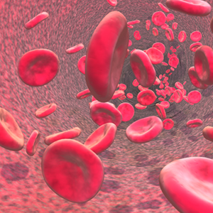
If you examine a stained smear of human blood cell under a microscope, you’ll find that there are different kind of cells, you will clearly see the red blood cells (the highest & clearest amount) scattered between them some white blood cells (having nuclei) & platelets, As just mentioned that the Erythrocytes vastly outnumber the other types of cells in the stained blood smear as seen in the following picture

THE ERYTHROCYTES
Structural Characteristics

Erythrocytes or red blood cells are small cells about 7.5 micro-meter in diameter, Having a shape like bioconcave discs–flattened discs with depressed or decreased centers, in which those thin centers appears lighter compared to the edges, all the mature erythrocytes are bounded together normally by a plasma membrane but lacking a nucleus as just mentioned & contains also no organelles, this can be clearly seen in the above pictures
They are basically like a box or reservoir of Hemoglobin (Hb) the RBC’s special protein used to transport gases, but still the RBC contains other kind of proteins which are used to maintain the plasma membrane & to promote changes in the RBC shape


An example for the other proteins helping in maintaing the structure of the RBC is the protein called "SPECTRIN" Which is forming normaly a network with other proteins to maintain the bioconcave structure of the RBC, it’s attached to cytoplasmic face of the RBC plasma membrane & because it is a flexible protein & deformable, so it do give the RBC structure some flexibility aiding it to turn, twist & become sometimes in a cup-shape while being carried along in the blood stream & through different sizes of different blood vessels & capillaries which sometimes might be smaller in diameter than the RBC it self, so RBC must be flexible & fits in different sizes before it can resume back it’s original bioconcave shape
The RBC’s are a very nice example of the complementary actions between the structure & the function, it fo picks up oxygen from the lungs, transport it to the other body cells than drop it there, in additon to that, it do also carries about 20% approximately of the wasted carbon dioxide from the body cells in the opposite direction carrying them back to the lungs capillaries, the structural characters of the RBC’s helps them to function in the proper way & do their job well because of the following
A) It’s small size & bioconcave shape provides a huge surface area compared to the volume, because of this shape it holds about 30% more surface area compared to a normal spherical cell, since no point in its cytoplasm is far from the surface, so it can be named as an ideal model for gas exchange process
B) Forgetting about the water content, the RBC is about 97% hemoglobin, which is the major functioning molecule in it
C) Because RBC’s lack cell organelles, especially there is no mitochondria, they do not consume any of the oxygen that they carry so that they are the best oxygen transporters compared to all the other cells, & they do generate energy by anaerobic mechanisms
Erythrocytes are the major reason of why the blood is VISCOUS, women normally have a lower RBC count compared to men, around (4.3 - 5.2 million cells per cubic millimeter mm3) of blood versus (5.1 - 5.8 million cells per cubic millimeter) respectively, And when the nuber of RBC increases cause of any reason the viscousity of the blood will increase & the blood flow will bcome slower in speed, after sometime the blood will re-thin again & flow normaly
THE ERYTHROCYTES FUNCTION
Erythrocytes were created in a very special way as mentioned before to be an ideal model for carrying gases (basically oxygen & nitrogen) from different locations to other locations in the body, this is it’s major function in the human body which is understood till now
They contain what is called "Hemoglobin" molecules which binds REVERSIBLY & easily with oxygen, in which most of the oxygen carried in the blood are bounded to those hemoglobin molecules, the normal values of hemoglobin are as the following
A) 14 to 20 g\100 ml of blood in infants
B) 13 to 18 g\100 ml in adult males
C) 13 to 16 g\100 ml in adult females
B) 13 to 18 g\100 ml in adult males
C) 13 to 16 g\100 ml in adult females
Hemoglobin Structure

This pictures above shows an oxygenated heme structure
Note that the coming pictures might vary in the chemical nature due to different ways of expressing the hemoglobin molecule & due to different chemical stages which the molecule might pass through while bounding & un-bounding to oxygen atoms, read further to understand the idea
Note that the coming pictures might vary in the chemical nature due to different ways of expressing the hemoglobin molecule & due to different chemical stages which the molecule might pass through while bounding & un-bounding to oxygen atoms, read further to understand the idea


The hemoglobin is made up of 2 basic elements first, a protein called Globin + A red Heme Pigment
The major hemoglobin in adults is called Hemoglobin A
The Globin Protein is composed of 4 poly-peptide chains, 2 of them are alpha & the other 2 are beta (as shown in the above pictures), in which each one of those poly-peptide chains binds to one heme group, so each globin protein binds basically to 4 heme groups, the heme group itself looks like a jewel in the middle of each poly-peptide chain of the 4 chains available in the globin protein molecule, in another words we can say that Hemoglobin A is composed of 2 dimers (alpha-beta 1) & (alpha-beta 2) where the numbers refers to dimer 1 & dimer 2, each dimer is holded tighlt by hydrophobic bonds, also the hydrophobic amino acid residues are not only localized internally, but also are available in a region outside on the surface of the subunit, so ionic & hydrogen bonds in addition to the another non-covalent bonds also occurs, all those different kinds of bonding between or inside the one dimer & between different dimers lead to the formation of a flexible but stable conformation structure which might change it’s conformation partially depending on the chemical situation, like when it binds oxygen it differs than when it binds 3 oxygen atoms & the same when those oxygen atoms dissociate or detach from the whole hemoglobin molecule
Each heme group is composed of an iron atom which is set in the center of the chemical formula as seen in the pictures above, each iron atom in a heme group can combine successfully & reversibly to one oxygen atom also, so each poly-peptide chain in the globin molecule can bind to one oxygen atom, in other way we can say that each hemoglobin molecule can bind to 4 oxygen atoms, because each hemoglobin contains 4 poly-peptide chains each one contains 1 heme group which binds to 1 oxygen atom
The Heme group origin of synthesis is based on basically 2 elements which are Glycine & Succinyl-Co-Enzyme A
The Heme-Group in the RBC is one kind of what is called HEMEPROTEINS which are available in more than one location in the human body, but each one holds a special function, the one in the RBC is for carrying oxygen & CO2, but the heme group of the cytochrome functions as an electron carrier that is altrenately oxidized & reduced, another example is the heme group of the enzyme CATALASE in which the heme group is considered a part of the active site of the enzyme that catalyzes the breakdown of hydrogen peroxide
المزيدThe blood Part 1
مايو 11th, 2009 كتبها اسماعيل العبد مرتضى نشر في , Human Anatomy & Physiology, The blood,Clinical Pharmacy, Human Anatomy & Physiology Section
The Blood Part 1
Studied & revised By Ismail Mortada
B.Sc.Pharmacy & Health Sciences, Clinical Pharmacy
دراسة الدم ومكوناته وخصائصه الفيزيائية, الكيميائية, التشريحية (لأنه يعتبر نسيج حي) والفيسيولوجية تعتبر من المواد المهمة جدا, نظرا لما يقوم به الدم من وظائف حيوية أساسية في جسم الانسان حيث أنه النهر الحي في جسمنا, ناقلا معه ما تيسر من مواد وتركيبات ووظائف أعجز شخصيا أنا أو غيري عن وصف أهميتها ومدى تعقيدها بشكل حصري وكامل ويحتاج الى مجلدات فعلا للتعمق في أموره وخصائصه, وتأتي دراسة وفهم فيسيولوجية وتشريح الدم صيدلانيا لأهميته القصوى في حركية الدواء في جسم الانسان ومدى تأثره سلبا وايجابا بوضع الدم بشكل عام, كما ان دراسته (بالاضافة الى باقي أجزاء الجسم) بشكل مفصل يعيننا على فهم أكثر تعمقا للطب والصيدلة بوجه عام
في هذا القسم الآن سنتحدث بشكل عام عن الدم ومكوناته العامة ونفصل قليلا في مكونات البلازما الدموية
في هذا القسم الآن سنتحدث بشكل عام عن الدم ومكوناته العامة ونفصل قليلا في مكونات البلازما الدموية
دراسة ومراجعة اسماعيل العبد المرتضى
يمكن تزويدكم بالمراجع المستخدمة تحت الطلب ويرجى الضغط على الصور لتكبيرها لقراءة محتوياتها
Blood was considered -and still considered- a vital very important system in the human body, it’s importancy was gained due to the varied functions done by the blood which are necessary for the human life & without it there would be no basic development of the human body, in which it serves as the life river in our bodies, this chain of lectures will study the blood from different points of view, because understanding the human anatomy & physiology including the blood studies are very necessary for pharmaceutical applications & for undertanding pharmacokinetics & Dynamics of the different drugs
Overview: Composition & functions of the Blood
Components


Blood is unique special anatomical structure & system in our body, it is a very special kind of Connective tissues & considered to be the only tissue in our body which appears in the liquid form, it’s composed of Living cells distributed in a non-living matrix (the plasma), & it doesn’t contain elastic & collagen fibers like the other tissues but still it contains dissolved fibrous proteins which becomes visible as Fibrin Strands when blood clotting occurs, the origin of the blood itself and it’s contents varies majorly between liver & other organs but basically the RBC’s are produced from the bone marrow
To check & study the blood direct visible contents we need to centrifuge it by placing a blood sample in a centrifuge machine & centrifuging it, so with the centrifugal force (gravity force) the heavy elements of the blood will accumulate down the centrifuge tube & the less heavy blood contents will flow at the top as shown in the above & below pictures


The above picture is for the machine used for blood centrifuging

We’ll find that the blood is composed of almost 3 basic visible layers arranged from up to down as the following
A) The upper layer composed of Plasma, which is about 55% of the blood volume
B) The thin whitish layer after the plasma called "Buffy Coat" which is composed of the White blood cells (Leukocytes) & the platelets which is about less than 1% of the blood volume
C) The Erythrocytes layer (Red Blood Cells), a layer red in colour cause it contains the red blood cells, which is about 45% of the blood volume
The red blood cells compose about 45% of the total volume of the blood giving a percentage known as Hematocrit, which is the percentage by volume of RBC’s in the blood, the normal values range between 47% (+5% or -5%) in normal healthy adult males upto 42% (+5% or -5%) in healthy adult females & the numbers will vary between different individuals in general, Blood plasma has a density of approximately 1025 kg/m3, or 1.025 kg/l
The blood is composed of a lot of different components other than the basic ingredients mentioned above, those components are distributed in the different layers & if we I want to be more specific, hundreds of real components will be mentioned
Blood Physical Characteristics & volume
Blood is a sticky opaque liquid with a well characterized metallic taste, the blood colour will vary widely depending on the amount of the oxygen carried by it ranging between dark red (oxygen-poor) up to scarlet lighter colour (oxygen-rich) blood, the blood is more denser than water & more viscous also due to the ingredients
The blood PH is slightly alkaline ranging between (7.35 upto 7.45) with a temperature about 38 degree’s centigrade or 100.4 degree fahrenheit which is always slightly higher than the normal body temperature
The blood PH is slightly alkaline ranging between (7.35 upto 7.45) with a temperature about 38 degree’s centigrade or 100.4 degree fahrenheit which is always slightly higher than the normal body temperature
BLOOD FUNCTIONS
Blood performs a number of functions, honestly all of them overlap & are complicated with each other forming the whole blood system, most of the blood functions includes the distribution of different substances between different locations, including also body regulation & protection functions, the blood itself plays an important role in controlling some natural physiological functions & is used also pharmaceutically in controlling the drugs pharmacokinetics, we can brief up the blood functions in general as the following
A) Distribution
B) Regulation
C) Protection
B) Regulation
C) Protection
A) Distribution
As well known, the blood functions as a transporter which aids different things to be re-located & distributed inside the human body including the following
A) Delivering oxygen from the lungs to all the body cells
B) Delivering nutrients (including any drugs, toxins or anything entering our body) from digestive system or skin or many method of body entrance to all the body cells
C) Transporting metabolic waste products from all the body cells to elimination sites, for example to the lungs for eliminating carbon dioxide & to the kidneys for disposal of nitrogenous & other wastes including the skin to eliminate sweat….etc
D) Transporting hormones from the Endocrine system & glands to their target organs
E) Holding & trapping all the blood ingredients in general from every thing including proteins, plasma ingredients & different blood formed elements such as RBC’s & WBC’s
In addition to the past mentioned natural physiological functions, I do add that the blood is important for distributing different chemicals (drugs & toxins) given to the human body so that they can reach their target cells, tissues & organs, this is very important in what is called Pharmacokinetics (which studies the drug distribution as a part of it) & it have an important applications -major applications- in pharmaceutical field, because any drug must reach the blood first so that we can say that it’s bioavailable than to reach a target location to exert a pharmacological action, blood contents & physiological state varies a lot individually & will affect the whole pharmacokinetics process in general
B) RegulationThe blood holds a very important responsibility in regulating a lot of natural physiological functions in the human body including the following
A) Maintaining appropriate body temperature by absorbing & distributing heat throughout the body & to the skin surface, encouraging heat loss & gain depending on the enviromental & physioloigcal states of the body & outside enviroment
B
ليست هناك تعليقات:
إرسال تعليق