
The Cardiovascular system, Blood Vessels Anatomy Part 4
Systemic Circulation
THE CAROTID ARTERIES
Specialized Lecture in Anatomy & Physiology
مناقشة إسماعيل مرتضى Discussed by Ismail Mortada
Systemic Circulation
THE CAROTID ARTERIES
Specialized Lecture in Anatomy & Physiology
مناقشة إسماعيل مرتضى Discussed by Ismail Mortada

Please press on the pictures to be able to see them in larger sizes so that to read the contents
As mentioned in past lectures, while we were discussing the systemic ciculation & blood vessels types, we said that near the aortic arch there are some branches of it, including the brachicephalic artery which raises both the right subclavian artery & the right common carotid artery + the other 2 branches from the aortic arch which raises the Left Common Carotid Artery & the Left Subclavian Artery
Now we’re going to discuss in more details about the common carotid arteries, now as just mentioned that there are 2 basic branches of the carotid artery which are as the following
A) The Right Common Carotid Artery which raise up from the Brachicephalic Artery from the Aorta Arch
B) The Left Common Carotid Artery which is one of the 3 branches of the Aorta Arch itself
A) The Right Common Carotid Artery which raise up from the Brachicephalic Artery from the Aorta Arch
B) The Left Common Carotid Artery which is one of the 3 branches of the Aorta Arch itself

They ascend through the lateran neck & at the superior border of the Larynx "at the level of ADAMS’ APPLE" & each one of the past mentioned 2 common carotid arteries divides into 2 major branches which are Internal & External
THE EXTERNAL CAROTID ARTERIES

It supply most tissues of the head except the brain & the orbit, as each artery runs superiorly it give the following branches
A) Superior Thyroid Artery , which supply the larynx & the thyroid gland
B) Lingual Artery , which supplies the tongue
C) Facial Artery , which supplies the skin & the muscles of the anterior face
D) Occipital Artery , which supplies the posterior scalp
A) Superior Thyroid Artery , which supply the larynx & the thyroid gland
B) Lingual Artery , which supplies the tongue
C) Facial Artery , which supplies the skin & the muscles of the anterior face
D) Occipital Artery , which supplies the posterior scalp


Each external carotid artery terminates by splitting into 2 endings which are as the following
A) Superficial Temporal Artery, which supplies the parotid salivary gland & most of the scalp
B) Maxillary Artery, which supplies the upper & lower jaws & chewing muscles, the teeth & the nazal cavities
A) Superficial Temporal Artery, which supplies the parotid salivary gland & most of the scalp
B) Maxillary Artery, which supplies the upper & lower jaws & chewing muscles, the teeth & the nazal cavities


From the Maxillary artery arises a clinically important artery called "THE MIDDLE MENINGEAL ARTERY" which enters the skull through the foramen spinosum & supplies the inner surface of the parietal bone, squamous region of the temporal bone & the underlying dura matter
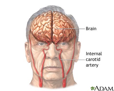
THE INTERNAL CAROTID ARTERY

THE AORTA
أكتوبر 31st, 2009 كتبها اسماعيل العبد مرتضى نشر في , Cardiovascular Ssytem, Human Anatomy & Physiology,The Cardiovascular system, Blood Vessels Anatomy Part 3
Systemic Circulation
THE AORTA
Specialized Lecture in Anatomy & Physiology
مناقشة إسماعيل مرتضى Discussed by Ismail Mortada
Systemic Circulation
THE AORTA
Specialized Lecture in Anatomy & Physiology
مناقشة إسماعيل مرتضى Discussed by Ismail Mortada

Please press on the pictures to be able to see them in larger sizes so that to read the contents
The Aorta is the largest blood vessel "Artery" in the human body, as it leaves the left ventricle of the heart carrying pure clean oxygenated blood to the other cells & tissues of the human body Its’ enternal diameter is about 2.5 cm & its’ wall is about 2 mm in thickness, the size of the aorta will continuously & slowly decrease in size while it runs to its terminus "the pelvix" as mentioned in lectures before
In addition, it contains a valve like in the heart, called "The Aortic Semilunar Valve" that protects the blood from going backward in its flush toward the tissues, opposite ot the semilunar valves is the AORTIC SINUS, which contains the BARORECEPTORS which are important for the reflex regulation of the blood pressure, each different portion of the aorta is named depending on its shape or location "as mentioned in past lectures the names reflects normaly shapes, locations of the bone nearby or even names of the organs being supported by the vessel itself", the different portions of the aorta starts with the "ASCENDING AORTA" which runs posteriorly & to the right side of the pulmonary trunk, it stays in this way for about 5 cm till it turns suddenly left near the aorta arch, after that there are branches which arise from this ascending aorta, the only basic 2 branches which are the RIGHT & LEFT CORONARY ARTERIES, which supply the mycardial muscle "the heart" it self
The aorta arches which is deep in the sternum begins & ends at the sternal angle (level of T4), its’ major branches which are right & left are as the following
A) The Brachiocephalic Artery "arm-head", which passes superiorly under the right clavicle and gives the rise to 2 more branches which are THE RIGHT COMMON CAROTID ARTERY & the THE RIGHT SUBCLAVIAN ARTERY
B) The Left Common Carotid Artery
C) The Left Subclavian Artery
B) The Left Common Carotid Artery
C) The Left Subclavian Artery


The past 3 mentioned vessels which arise from the aorta arches serves the head, neck, upper limbs & part of the Thorax wall
After we discussed the Ascending Aorta & the aorta arch with its 3 blood vessel branches we reach now to the DESCENDING AORTA or the THORACIC ARTERY which will runn from (T5 to T12) & in this trip it branches to several arteries which serve the thorax & viscera before it penetrates the diaphragm
After penetrating the diaphragm, the portion will turn to become the ABDOMINAL AORTA, in which it supplies the abdominal walls & viscera & ends at the level of L4 in which there it will split giving the RIGHT & LEFT COMMON ILIAC ARTERIES, which supplies the pelvis & the lower limbs

So as a brief for the parts mentioned before we see the following
A) The blood will leave the left ventricle via the aorta
B)
المزيدA) The blood will leave the left ventricle via the aorta
B)
Systemic Circulation
أكتوبر 30th, 2009 كتبها اسماعيل العبد مرتضى نشر في , Cardiovascular Ssytem, Human Anatomy & Physiology,The Cardiovascular system, Blood Vessels Anatomy Part II
Systemic Circulation
Specialized Lecture in Anatomy & Physiology
مناقشة إسماعيل مرتضى Discussed by Ismail Mortada
Systemic Circulation
Specialized Lecture in Anatomy & Physiology
مناقشة إسماعيل مرتضى Discussed by Ismail Mortada
The Major reference for the this lecture & the following lectures in the same subject is
"Human Anatomy & Physiology" For Elaine N.Marieb the 5′th Edition
+ Internet + My personal University Lecture Notes
"Human Anatomy & Physiology" For Elaine N.Marieb the 5′th Edition
+ Internet + My personal University Lecture Notes

Please press on the pictures to be able to see them in larger sizes so that to read the contents
As mentioned in the lecture part (I) when we spoke about the general anatomy of the blood vessels & we said that it is made up of 2 major sections which are the pulmonary circulation & the systemic circulation, we finished discussing in brief the pulmonary circulation before & we mentioned the different arteries & veins which were included in this part of the circulation & now before going on with the lectures speaking about the Systemic Circulation it is good to remind ourselves that in the systemic circulation "which will be discussed now" the arteries are the vessels carrying oxygenated blood from the heart to the other body cells & tissues & the veins are those vessels which carries the blood toward the heart which is poor in oxygen and more rich in waste materials & carbon dioxide
B) The Systemic Circulation

The Systemic circulation starts in the heart after receiving the clean oxygenated blood from the pulmonary arteries, the oxygenated blood will enter the LEFT HEART ATRIUM & will be pumped from there to the LEFT HEART VENTRICLE; & from there the journey will start through the AORTA, which is the basic & largest vessel "arterie" in the human body from there, from the aorta, the blood can move on in different pathways, since all the major blood vessels arise from this single large AORTA , It normally ARCHES upward from the heart & then curves & run down along the body midline in which it terminates in the PELVIS where there it splits forming the 2 large branches arteries that serve the lower extremities, then the branches of the arteries forms the smaller ARTERIOLES & then as usual forming the Blood Capillaries in which the exchange of gases & nutrients occurs on this level

So we can brief the past steps as the following
A) The blood will enter from the Pulmonary arteries to the Left Heart Atrium
B) From there it will be entering the Left Heart Ventricle to be pumped out to the Aorta
C) The Aorta is the major blood vessel "Arterie" which rises from the heart
D)
المزيدA) The blood will enter from the Pulmonary arteries to the Left Heart Atrium
B) From there it will be entering the Left Heart Ventricle to be pumped out to the Aorta
C) The Aorta is the major blood vessel "Arterie" which rises from the heart
D)
Pulmonary Circulation
أكتوبر 28th, 2009 كتبها اسماعيل العبد مرتضى نشر في , Cardiovascular Ssytem, Human Anatomy & Physiology,The Cardiovascular system, Blood Vessels Anatomy
Pulmonary Circulation
Specialized Lecture in Anatomy & Physiology
مناقشة إسماعيل مرتضى Discussed by Ismail Mortada
The Major reference for the this lecture & the following lectures in the same subject is
"Human Anatomy & Physiology" For Elaine N.Marieb the 5′th Edition
+ Internet + My personal University Lecture Notes
"Human Anatomy & Physiology" For Elaine N.Marieb the 5′th Edition
+ Internet + My personal University Lecture Notes

Please press on the pictures to be able to see them in larger sizes so that to read the contents
As it is known that the cardiovascular system is composed of 2 major components which are the Heart & the Vascular System it self "The Blood Vessels" in which they complete each other within the dynamic physiology in the human body & in other creatures also on this earth & my basic aim now in those lectures is to revise the names, locations & brief physiology of the blood vessels in the human body
Excluding the heart anatomy & physiology, it comes directly the vascular system which is made up of a blend of different kinds of arteries & veins which are used to transport different forms of the blood & nutrient from & to the heart & within the entire community of the human body cells , the circulation is made up of 2 basic sections which are as the following
A) The Pulmonary Circulation
B) The Systemic Circulation
A) The Pulmonary Circulation
B) The Systemic Circulation
A) The Pulmonary Circulation

Now, Considering the blood which is poor in oxygen & coming from all over the body cells & tissues toward the heart, it will enter the RIGHT ATRIUM of the heart & from it, it will be pumped to the RIGHT VENTRICLE & from there it will be sent into what is called the "Pulmonary Trunk", which runs diagonally upward for about 8 cm & then will be divided into the RIGHT & LEFT PULMONARY ARTERIES; Further to that, those pulmonary arteries will be divided inside the lungs to form the Lobar arteries in which there are 3 lobar arteries in the right lung & 2 lobar arteries in the left lung each one of the general 5 lobar arteries serves one lung lobe respectively, from the lobar arteries it branches to form the ARTERIOLES & then finally to give the PULMONARY CAPILLARIES that surround the air sacs & it is there in the smallest capillaries where the gases exchange will start in the human body

Note that people are used to use the word arterie for the blood vessel which carries pure oxygenated blood, but this is not the case here neither it is the right description for what’s exactly going on in anatomy & physiology, since in the systemic circulation, arterie would be any vessel which carries blood from the heart to any cell or tissue in the human body from which also is the lungs, so the blood entering the lungs "in the pulmonary circulation" coming from the tissues and poorly oxygenated is still called arterie and the blood leaving the lungs to the heart is called the veins, so in the pulmonary circulation the veins are the one who carries the oxygenated blood toward the left heart atrium & the arteries are those going out from the right heart ventricle toward the lungs, this is an important note in anatomy & physiology to be always considered & in addition to that any vessel which contains in its name the word "Pulmonary" or "Lobar" means that it belongs to the Pulmonary Circulation or the lungs & doesn’t belong to the Systemic Circulation


So as a faster brief to the mentioned information below we can say the following
A) The poor oxygenated blood will enter the Right lobe of the heart to the right atrium
B) Then it will be sent or pumped from the right heart atrium to the right heart ventricle
C) Then it will be pumped to the Pulmonary Trunk
D) From the pulmonary trunk it will be sent to both the left & the right pulmonary arteries
E) The pulmonary arterie will form what is called the Lobar Arteries in which they are basically 5 in numbers
F) The lobar arteries are 3 arteries in the right lungs serving 3 lobes
G) The lobar arteries are forming 2 arteries in the left lung serving the lobes there aloso & the totall as mentioned before are 5 alobar arteries
H) Then the lobar arteries will divide forming smaller Arterioles
I) Then the arterioles will form the smalles Pulmonary Capillaries where the gas exchange will occur


In the alveoli or the air sacs in the lungs, the oxygen exchange will occur & the blood will become brighter red in color "due to the hemoglobin change & iron state change & oxygen availability" so in this case the blood will keep on moving from the pulmonary capillaries into the VENULES, from which it will form the PULMONARY VEINS exiting from each lung lobe, so in total there are 4 pulmonary veins arising from the lungs in general all of which moves toward the left heart atrium
Please watch the following videos which are related in a way or other to the subject of this lecture
r
ليست هناك تعليقات:
إرسال تعليق