
The Cardiovascular system, Blood Vessels Anatomy Part 4
Systemic Circulation
THE CAROTID ARTERIES
Specialized Lecture in Anatomy & Physiology
مناقشة إسماعيل مرتضى Discussed by Ismail Mortada
Systemic Circulation
THE CAROTID ARTERIES
Specialized Lecture in Anatomy & Physiology
مناقشة إسماعيل مرتضى Discussed by Ismail Mortada

Please press on the pictures to be able to see them in larger sizes so that to read the contents
As mentioned in past lectures, while we were discussing the systemic ciculation & blood vessels types, we said that near the aortic arch there are some branches of it, including the brachicephalic artery which raises both the right subclavian artery & the right common carotid artery + the other 2 branches from the aortic arch which raises the Left Common Carotid Artery & the Left Subclavian Artery
Now we’re going to discuss in more details about the common carotid arteries, now as just mentioned that there are 2 basic branches of the carotid artery which are as the following
A) The Right Common Carotid Artery which raise up from the Brachicephalic Artery from the Aorta Arch
B) The Left Common Carotid Artery which is one of the 3 branches of the Aorta Arch itself
A) The Right Common Carotid Artery which raise up from the Brachicephalic Artery from the Aorta Arch
B) The Left Common Carotid Artery which is one of the 3 branches of the Aorta Arch itself

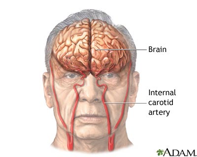
They ascend through the lateran neck & at the superior border of the Larynx "at the level of ADAMS’ APPLE" & each one of the past mentioned 2 common carotid arteries divides into 2 major branches which are Internal & External
THE EXTERNAL CAROTID ARTERIES

It supply most tissues of the head except the brain & the orbit, as each artery runs superiorly it give the following branches
A) Superior Thyroid Artery , which supply the larynx & the thyroid gland
B) Lingual Artery , which supplies the tongue
C) Facial Artery , which supplies the skin & the muscles of the anterior face
D) Occipital Artery , which supplies the posterior scalp
A) Superior Thyroid Artery , which supply the larynx & the thyroid gland
B) Lingual Artery , which supplies the tongue
C) Facial Artery , which supplies the skin & the muscles of the anterior face
D) Occipital Artery , which supplies the posterior scalp


Each external carotid artery terminates by splitting into 2 endings which are as the following
A) Superficial Temporal Artery, which supplies the parotid salivary gland & most of the scalp
B) Maxillary Artery, which supplies the upper & lower jaws & chewing muscles, the teeth & the nazal cavities
A) Superficial Temporal Artery, which supplies the parotid salivary gland & most of the scalp
B) Maxillary Artery, which supplies the upper & lower jaws & chewing muscles, the teeth & the nazal cavities


From the Maxillary artery arises a clinically important artery called "THE MIDDLE MENINGEAL ARTERY" which enters the skull through the foramen spinosum & supplies the inner surface of the parietal bone, squamous region of the temporal bone & the underlying dura matter
THE INTERNAL CAROTID ARTERY

THE AORTA
أكتوبر 31st, 2009 كتبها اسماعيل العبد مرتضى نشر في , Cardiovascular Ssytem, Human Anatomy & Physiology,The Cardiovascular system, Blood Vessels Anatomy Part 3
Systemic Circulation
THE AORTA
Specialized Lecture in Anatomy & Physiology
مناقشة إسماعيل مرتضى Discussed by Ismail Mortada
Systemic Circulation
THE AORTA
Specialized Lecture in Anatomy & Physiology
مناقشة إسماعيل مرتضى Discussed by Ismail Mortada

Please press on the pictures to be able to see them in larger sizes so that to read the contents
The Aorta is the largest blood vessel "Artery" in the human body, as it leaves the left ventricle of the heart carrying pure clean oxygenated blood to the other cells & tissues of the human body Its’ enternal diameter is about 2.5 cm & its’ wall is about 2 mm in thickness, the size of the aorta will continuously & slowly decrease in size while it runs to its terminus "the pelvix" as mentioned in lectures before
In addition, it contains a valve like in the heart, called "The Aortic Semilunar Valve" that protects the blood from going backward in its flush toward the tissues, opposite ot the semilunar valves is the AORTIC SINUS, which contains the BARORECEPTORS which are important for the reflex regulation of the blood pressure, each different portion of the aorta is named depending on its shape or location "as mentioned in past lectures the names reflects normaly shapes, locations of the bone nearby or even names of the organs being supported by the vessel itself", the different portions of the aorta starts with the "ASCENDING AORTA" which runs posteriorly & to the right side of the pulmonary trunk, it stays in this way for about 5 cm till it turns suddenly left near the aorta arch, after that there are branches which arise from this ascending aorta, the only basic 2 branches which are the RIGHT & LEFT CORONARY ARTERIES, which supply the mycardial muscle "the heart" it self
The aorta arches which is deep in the sternum begins & ends at the sternal angle (level of T4), its’ major branches which are right & left are as the following
A) The Brachiocephalic Artery "arm-head", which passes superiorly under the right clavicle and gives the rise to 2 more branches which are THE RIGHT COMMON CAROTID ARTERY & the THE RIGHT SUBCLAVIAN ARTERY
B) The Left Common Carotid Artery
C) The Left Subclavian Artery
B) The Left Common Carotid Artery
C) The Left Subclavian Artery


The past 3 mentioned vessels which arise from the aorta arches serves the head, neck, upper limbs & part of the Thorax wall
After we discussed the Ascending Aorta & the aorta arch with its 3 blood vessel branches we reach now to the DESCENDING AORTA or the THORACIC ARTERY which will runn from (T5 to T12) & in this trip it branches to several arteries which serve the thorax & viscera before it penetrates the diaphragm
After penetrating the diaphragm, the portion will turn to become the ABDOMINAL AORTA, in which it supplies the abdominal walls & viscera & ends at the level of L4 in which there it will split giving the RIGHT & LEFT COMMON ILIAC ARTERIES, which supplies the pelvis & the lower limbs

So as a brief for the parts mentioned before we see the following
A) The blood will leave the left ventricle via the aorta
B)
المزيدA) The blood will leave the left ventricle via the aorta
B)
Systemic Circulation
أكتوبر 30th, 2009 كتبها اسماعيل العبد مرتضى نشر في , Cardiovascular Ssytem, Human Anatomy & Physiology,The Cardiovascular system, Blood Vessels Anatomy Part II
Systemic Circulation
Specialized Lecture in Anatomy & Physiology
مناقشة إسماعيل مرتضى Discussed by Ismail Mortada
Systemic Circulation
Specialized Lecture in Anatomy & Physiology
مناقشة إسماعيل مرتضى Discussed by Ismail Mortada
The Major reference for the this lecture & the following lectures in the same subject is
"Human Anatomy & Physiology" For Elaine N.Marieb the 5′th Edition
+ Internet + My personal University Lecture Notes
"Human Anatomy & Physiology" For Elaine N.Marieb the 5′th Edition
+ Internet + My personal University Lecture Notes

Please press on the pictures to be able to see them in larger sizes so that to read the contents
As mentioned in the lecture part (I) when we spoke about the general anatomy of the blood vessels & we said that it is made up of 2 major sections which are the pulmonary circulation & the systemic circulation, we finished discussing in brief the pulmonary circulation before & we mentioned the different arteries & veins which were included in this part of the circulation & now before going on with the lectures speaking about the Systemic Circulation it is good to remind ourselves that in the systemic circulation "which will be discussed now" the arteries are the vessels carrying oxygenated blood from the heart to the other body cells & tissues & the veins are those vessels which carries the blood toward the heart which is poor in oxygen and more rich in waste materials & carbon dioxide
B) The Systemic Circulation

The Systemic circulation starts in the heart after receiving the clean oxygenated blood from the pulmonary arteries, the oxygenated blood will enter the LEFT HEART ATRIUM & will be pumped from there to the LEFT HEART VENTRICLE; & from there the journey will start through the AORTA, which is the basic & largest vessel "arterie" in the human body from there, from the aorta, the blood can move on in different pathways, since all the major blood vessels arise from this single large AORTA , It normally ARCHES upward from the heart & then curves & run down along the body midline in which it terminates in the PELVIS where there it splits forming the 2 large branches arteries that serve the lower extremities, then the branches of the arteries forms the smaller ARTERIOLES & then as usual forming the Blood Capillaries in which the exchange of gases & nutrients occurs on this level

So we can brief the past steps as the following
A) The blood will enter from the Pulmonary arteries to the Left Heart Atrium
B) From there it will be entering the Left Heart Ventricle to be pumped out to the Aorta
C) The Aorta is the major blood vessel "Arterie" which rises from the heart
D)
المزيدA) The blood will enter from the Pulmonary arteries to the Left Heart Atrium
B) From there it will be entering the Left Heart Ventricle to be pumped out to the Aorta
C) The Aorta is the major blood vessel "Arterie" which rises from the heart
D)
Pulmonary Circulation
أكتوبر 28th, 2009 كتبها اسماعيل العبد مرتضى نشر في , Cardiovascular Ssytem, Human Anatomy & Physiology,The Cardiovascular system, Blood Vessels Anatomy
Pulmonary Circulation
Specialized Lecture in Anatomy & Physiology
مناقشة إسماعيل مرتضى Discussed by Ismail Mortada
The Major reference for the this lecture & the following lectures in the same subject is
"Human Anatomy & Physiology" For Elaine N.Marieb the 5′th Edition
+ Internet + My personal University Lecture Notes
"Human Anatomy & Physiology" For Elaine N.Marieb the 5′th Edition
+ Internet + My personal University Lecture Notes

Please press on the pictures to be able to see them in larger sizes so that to read the contents
As it is known that the cardiovascular system is composed of 2 major components which are the Heart & the Vascular System it self "The Blood Vessels" in which they complete each other within the dynamic physiology in the human body & in other creatures also on this earth & my basic aim now in those lectures is to revise the names, locations & brief physiology of the blood vessels in the human body
Excluding the heart anatomy & physiology, it comes directly the vascular system which is made up of a blend of different kinds of arteries & veins which are used to transport different forms of the blood & nutrient from & to the heart & within the entire community of the human body cells , the circulation is made up of 2 basic sections which are as the following
A) The Pulmonary Circulation
B) The Systemic Circulation
A) The Pulmonary Circulation
B) The Systemic Circulation
A) The Pulmonary Circulation

Now, Considering the blood which is poor in oxygen & coming from all over the body cells & tissues toward the heart, it will enter the RIGHT ATRIUM of the heart & from it, it will be pumped to the RIGHT VENTRICLE & from there it will be sent into what is called the "Pulmonary Trunk", which runs diagonally upward for about 8 cm & then will be divided into the RIGHT & LEFT PULMONARY ARTERIES; Further to that, those pulmonary arteries will be divided inside the lungs to form the Lobar arteries in which there are 3 lobar arteries in the right lung & 2 lobar arteries in the left lung each one of the general 5 lobar arteries serves one lung lobe respectively, from the lobar arteries it branches to form the ARTERIOLES & then finally to give the PULMONARY CAPILLARIES that surround the air sacs & it is there in the smallest capillaries where the gases exchange will start in the human body

Note that people are used to use the word arterie for the blood vessel which carries pure oxygenated blood, but this is not the case here neither it is the right description for what’s exactly going on in anatomy & physiology, since in the systemic circulation, arterie would be any vessel which carries blood from the heart to any cell or tissue in the human body from which also is the lungs, so the blood entering the lungs "in the pulmonary circulation" coming from the tissues and poorly oxygenated is still called arterie and the blood leaving the lungs to the heart is called the veins, so in the pulmonary circulation the veins are the one who carries the oxygenated blood toward the left heart atrium & the ar
المزيدErythrocytes Production
أغسطس 28th, 2009 كتبها اسماعيل العبد مرتضى نشر في , Human Anatomy & Physiology, The blood,Clinical Pharmacy, Human Anatomy & Physiology Section
The Blood Part 3, Erythrocytes Production
Studied & revised By Ismail Mortada
B.Sc.Pharmacy & Health Sciences
MBA; Pharmaceutical Marketing Masters; Under Process
MBA; Pharmaceutical Marketing Masters; Under Process
دراسة الدم ومكوناته وخصائصه الفيزيائية, الكيميائية, التشريحية (لأنه يعتبر نسيج حي) والفيسيولوجية تعتبر من المواد المهمة جدا, نظرا لما يقوم به الدم من وظائف حيوية أساسية في جسم الانسان حيث أنه النهر الحي في جسمنا, ناقلا معه ما تيسر من مواد وتركيبات ووظائف أعجز شخصيا أنا أو غيري عن وصف أهميتها ومدى تعقيدها بشكل حصري وكامل ويحتاج الى مجلدات فعلا للتعمق في أموره وخصائصه, وتأتي دراسة وفهم فيسيولوجية وتشريح الدم صيدلانيا لأهميته القصوى في حركية الدواء في جسم الانسان ومدى تأثره سلبا وايجابا بوضع الدم بشكل عام, كما ان دراسته (بالاضافة الى باقي أجزاء الجسم) بشكل مفصل يعيننا على فهم أكثر تعمقا للطب والصيدلة بوجه عام
في هذا القسم الآن سنتحدث بشكل أكثر تخصصا ومتابعة للمقالات السابقة حول طريقة ولادة وتكون كريات الدم الحمراء من نخاع العظم, وآلية التكوين وخطواته المتتالية وأهمية ذلك وكيفية تأثيره على ظهور بعض الأمراض في بعض الأحيان
في هذا القسم الآن سنتحدث بشكل أكثر تخصصا ومتابعة للمقالات السابقة حول طريقة ولادة وتكون كريات الدم الحمراء من نخاع العظم, وآلية التكوين وخطواته المتتالية وأهمية ذلك وكيفية تأثيره على ظهور بعض الأمراض في بعض الأحيان
دراسة ومراجعة اسماعيل العبد مرتضى
يمكن تزويدكم بالمراجع المستخدمة تحت الطلب ويرجى الضغط على الصور لتكبيرها لقراءة محتوياتها

In last lectures, we spoke about the blood in general & about the RBC’s, which are the Red Blood Cells, in this part we’re going to go more deeper in discussing the production process of the RBC’s which is called "Hematopoiesis", or also called "Hemopoiesis"; and this is the major process to get a new born or new made red blood cells
The Bone Marrow

The bone marrow briefly is the location were RBC’s are made; it is composed basically of a large soft network of Reticular Connective Tissue that borders on wide blood capillaries called "Blood Sinusoids". Within this network there are some of the following elements
A) Immature blood cells
B) Macrophages
C) Fat cells
D) Reticular cells; which are the fibroblasts that secrets the fibers
B) Macrophages
C) Fat cells
D) Reticular cells; which are the fibroblasts that secrets the fibers
As it is defined normally that there are 2 type of bone marrows; one is the Red Bone Marrow & the other is the Yellow Bone Marrow, both have similar characteristics in which the red one is consisting mainly of Myeloid Tissues & the yellow one is consisting mainly of fat cells; but now we are concerned more about the Red Bone Marrow as it is the place were the RBC’S, WBC’S & platelets are formed & manufactured, In adults, Red Bone Marrow is found chiefly in the bones of the axial skeleton & gridles, & in the proximal epiphyses of the humerus & femur
Each type of blood cell is produced in different numbers in response to changing body needs & different regulatory factors. As they mature, they migrate through the thin walls of the sinusoids to enter the blood stream. On average, the marrow turns out an ounce of new blood containing around 100 billion new cells each & every day
Although the different produced elements and cells, each have specific functions, but still there are similarities occuring between all of them, Now all the elements arise from the same STEM CELL, the pleuripotential hematopoietic stem cell, or what is also called hemocytoblast, which resides in the red bone marrow, now although they all arise from the same origin but still their maturation process & pathway is different, & once a cell went within a special maturation pathway, it cannot after that change the way back, This commitment is signaled by the appearance of Membrane Surface Receptors that respond only to specific hormones & growth factors, which at the end activate the cell toward a further specific specialization
Erythropoiesis

The Erythrocyte production "Erythropoiesis" begins when a hemocytoblast descendant called a Myeloid Stem Cell is transformed into proerythroblast, which in turn gives arise to the early (basophilic) erythroblasts that produce huge numbers of ribosomes, During these first 2 phases, the cells divide many times. The Hemoglobin synthesis & iron accumulation occurs as the early erythroblast is transformed into a Late Erythroblast. The color of the cell cytoblasm changes as the Blue-Staining Ribosomes becomes masked by the pink color of Hemoglobin, In this stage & when the last cell produced "Normoblast" accumulates hemoglobin in a concentration of around 34%, most of its organelles are ejected, In addition, it’s nuclear functions ends & is pinched off. This will lead to cell collapsion "To Inward Direction" & will take at the end the bioconcave shape, so the result after assuming the bioconcave shape will be the what is called "The Reticulocyte" , those reticulocytes are considered to be the young erythrocytes which are holding that name because they still contain a network of clumped ribosomes & rough endoplasmic reticulum. All the entire process from forming the hemocytoblast untill forming the reticulocyte will take around 3 to 5 days, The filled reticulocytes almost are filled to bursting with hemoglobin, then those reticulocytes filled with hemoglobin will enter the blood stream to begin their functions & tasks in oxygen transport. Usually they become fully matured eryhthrocytes whithin 2 days of the release of the reticulocytes, as the reticulocytes ribosomes are degraded by intracellular enzymes
Please check the next picture which shows the exact conversion process starting from the hemoblast & ending up with the erythrocyte

Please press on the picture to increase the size & clarify the contents
Please press on the picture to increase the size & clarify the contents
So as a brief to the erythropiesis steps, it would be in a more organized way as the following
A) When the Myeloid Stem cell is transformed into Proerythrocyte
B) Then this proerythrocyte is transformed into a basophilic erythroblast, in this step a huge numer of ribosomes are produced and Blue color is the major color of the cells till now & in the steps A & B the cells divide so many times in a fast way
C) Then the proerythrocyte will become an Early erythroblast & this is when also at the same time iron accumulation & hemoglobin synthesis will start
D) In step C also the color will change to become pink & this is after the accumulation of hemoglobin
E) Then the normoblast is formed from the late erythroblast, the normoblast will acuumulate hemoglobin in good concentration & will eject some cell organelles & will become in a bioconcave shape also
F) This bioconcaved shape cell will become a reticulocyte which is considered to be the young erythrocyte
G) The whole past steps will take around 3 to 5 days
H) The reticulocyte will be released to the blood & within 2 days after degrading the ribosomes it will become a mature red blood cell
The reticulocytes accounts for 1-2% of all the erythrocytes in the blood of healthy people, This Reticulocytes Count is used clinically as a rough index of the rate of the RBCs’ formation, so the if the count was below or above that range, this will indicates an ubnormal rates of erythrocyte formation

You need to keep in mind always different factors when studying the erythropoiesis process & although they show to be not important for some students but I see my self that they are very essential & critical for understanding the whole mechanism of the RBC formation, you need to check & understand the time taken for each step & the colors transformation, because all this will help future diagnosig of problems & further studies & researches for the blood, also you need to keep in count the size of the cells in the different levels & the organelles included in each step
المزيدالإسهال بدون طبيب..منقول عن مدونة الفيل
أغسطس 24th, 2009 كتبها اسماعيل العبد مرتضى نشر في , Digestive system, General Medical Articles, Human Anatomy & Physiology,نقلا عن مدونة الفيل-النت بتتكلم عربي
أحببت ان أنقل هذه المقالة السريعة وهي متعلقة بموضوع الإسهال والقيء والخ..وجدت أنها سهلة وممتنعة وبلغة قريبة
نقلت عن مدونة الفيل بالعنوان الموجود أسفل هذا السطر
تحياتي
إسماعيل مرتضى
hadoota.maktoobblog.com/1612815/%D8%A7%D9%84%D8%A7%D8%B3%D9%87%D8%A7%D9%84-%D8%A8%D8%AF%D9%88%D9%86-%D8%B7%D8%A8%D9%8A%D8%A8/#comment-2556517كتبهاالفيل–النت بتتكلم عربى ، في 23 أغسطس 2009 الساعة: 22:49 م
الاسها ل هو زيادة عدد مرات التبرز عن المعتاد للشخص (اكتر من 3 مرات يوميا) او زيادة سيلان قوام البراز عن المعتاد بالنسبة للشخصوالاسهال دائما ما ينتج عن خلل فى التوازن الطبيعى -والخلل فى الميكانزمات الطبيعية يعنى المرض وفى حالة الاسهال سيكون سبب الخلل هو عنصر طارئ(ميكروب مثلا- تناوله الانسان فى طعامه او شراب ملوث
وفى كل الاحوال فان الاسهال ينتج عن ذيادة محتوى الماء-والاملاح- فى البراز كفاقد وهى ماكان يجب على الجسم ان يحتفظ باغلبها نظرا لخلل فى ميكانيزمات امتصاصها-او الاحتفاظ بها-ا بالامعاء والقولون نتيجة لعامل طارئ كميكروب اومادة اخرى تخل بتوازن ميكانزمات الامتصاص فى الامعاء
وفى اغلب الاحوال لايتعدى اثر الاسهال عن كونة خلل مؤقت فاغلب حالات الاسهال هى حالات محدودة بذاتها وتنهى نفسها بنفسها
فالاسهال هو اسلوب دفاعى فريد حيث يقوم الجسم بتخفيف وطرد الاذى الطارئ فى الامعاء(ميكروب مثلا)
وهو يماثل القئ كاسلوب دفاعى اذا ماكان الميكروب او المادة الطارئة فى الجهاز الهضمى العلوى(المعدة) او الكحة اذا ماكانت المشكلة ف الجهاز التنفسى
كل هذة الميكانزمات هى وسائل تسعى الى تخفيف وطرد والتخلص من سبب الخلل بطرده خارج الجسم
واعاقتها وكبتها يعنى الاحتفاظ بالاذى داخل الجسم وهو غير مستحب اطلاقا كما فى حالة ادوية الاسهال_ او ادوية مضادات المغص-(بسكوبان .. الخ) التى توقف الحركة الطاردة للامعاء(الاسهال كدفاع*)
لذلك فاى علاج رشيد لايجب ان يسعى الى كبت وتعطيل الوسائل الدفاعية الطبيعية كالاسهال او القئ او الكحة بقدر ما يجب ان يرك
المزيد
The Blood Part 2, Erythrocytes
مايو 13th, 2009 كتبها اسماعيل العبد مرتضى نشر في , Human Anatomy & Physiology, The blood,Clinical Pharmacy, Human Anatomy & Physiology Section
The Blood Part 2, Erythrocytes
Studied & revised By Ismail Mortada
B.Sc.Pharmacy & Health Sciences, Clinical Pharmacy
دراسة الدم ومكوناته وخصائصه الفيزيائية, الكيميائية, التشريحية (لأنه يعتبر نسيج حي) والفيسيولوجية تعتبر من المواد المهمة جدا, نظرا لما يقوم به الدم من وظائف حيوية أساسية في جسم الانسان حيث أنه النهر الحي في جسمنا, ناقلا معه ما تيسر من مواد وتركيبات ووظائف أعجز شخصيا أنا أو غيري عن وصف أهميتها ومدى تعقيدها بشكل حصري وكامل ويحتاج الى مجلدات فعلا للتعمق في أموره وخصائصه, وتأتي دراسة وفهم فيسيولوجية وتشريح الدم صيدلانيا لأهميته القصوى في حركية الدواء في جسم الانسان ومدى تأثره سلبا وايجابا بوضع الدم بشكل عام, كما ان دراسته (بالاضافة الى باقي أجزاء الجسم) بشكل مفصل يعيننا على فهم أكثر تعمقا للطب والصيدلة بوجه عام
في هذا القسم الآن سنتحدث بشكل عام عن الدم ومكوناته العامة وخصوصا كريات الدم الحمراء
في هذا القسم الآن سنتحدث بشكل عام عن الدم ومكوناته العامة وخصوصا كريات الدم الحمراء
دراسة ومراجعة اسماعيل العبد المرتضى
يمكن تزويدكم بالمراجع المستخدمة تحت الطلب ويرجى الضغط على الصور لتكبيرها لقراءة محتوياتها

The Formed Elements
The formed elements of the blood (other than the plasma & the other contents mentioned in past articles) includes the following
A) The Erythrocytes, called Red Blood Cells or RBC’s
B) The Leukocytes, called White Blood Cells or WBC’s
C) The Platelets
A) The Erythrocytes, called Red Blood Cells or RBC’s
B) The Leukocytes, called White Blood Cells or WBC’s
C) The Platelets
Those formed elements have a very special characters, characterizing them from other kind of cells which are as the following
A) 2 of the 3 types of contents are not even true cells, the RBC’s doesn’t have a nuclei or organelles & the platelets are just simply a cell fragments
B) Only the Leukocytes are considered to be a complete cells
C) Most of the formed elements survive in the blood for only few days
D) Most blood cells do not devide, instead, they are continously renewed by division of cells in the bone marrow, where they originate
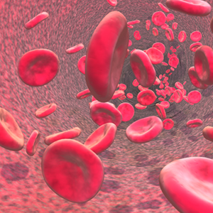
If you examine a stained smear of human blood cell under a microscope, you’ll find that there are different kind of cells, you will clearly see the red blood cells (the highest & clearest amount) scattered between them some white blood cells (having nuclei) & platelets, As just mentioned that the Erythrocytes vastly outnumber the other types of cells in the stained blood smear as seen in the following picture

THE ERYTHROCYTES
Structural Characteristics

Erythrocytes or red blood cells are small cells about 7.5 micro-meter in diameter, Having a shape like bioconcave discs–flattened discs with depressed or decreased centers, in which those thin centers appears lighter compared to the edges, all the mature erythrocytes are bounded together normally by a plasma membrane but lacking a nucleus as just mentioned & contains also no organelles, this can be clearly seen in the above pictures
They are basically like a box or reservoir of Hemoglobin (Hb) the RBC’s special protein used to transport gases, but still the RBC contains other kind of proteins which are used to maintain the plasma membrane & to promote changes in the RBC shape


An example for the other proteins helping in maintaing the structure of the RBC is the protein called "SPECTRIN" Which is forming normaly a network with other proteins to maintain the bioconcave structure of the RBC, it’s attached to cytoplasmic face of the RBC plasma membrane & because it is a flexible protein & deformable, so it do give the RBC structure some flexibility aiding it to turn, twist & become sometimes in a cup-shape while being carried along in the blood stream & through different sizes of different blood vessels & capillaries which sometimes might be smaller in diameter than the RBC it self, so RBC must be flexible & fits in different sizes before it can resume back it’s original bioconcave shape
The RBC’s are a very nice example of the complementary actions between the structure & the function, it fo picks up oxygen from the lungs, transport it to the other body cells than drop it there, in additon to that, it do also carries about 20% approximately of the wasted carbon dioxide from the body cells in the opposite direction carrying them back to the lungs capillaries, the structural characters of the RBC’s helps them to function in the proper way & do their job well because of the following
A) It’s small size & bioconcave shape provides a huge surface area compared to the volume, because of this shape it holds about 30% more surface area compared to a normal spherical cell, since no point in its cytoplasm is far from the surface, so it can be named as an ideal model for gas exchange process
B) Forgetting about the water content, the RBC is about 97% hemoglobin, which is the major functioning molecule in it
C) Because RBC’s lack cell organelles, especially there is no mitochondria, they do not consume any of the oxygen that they carry so that they are the best oxygen transporters compared to all the other cells, & they do generate energy by anaerobic mechanisms
Erythrocytes are the major reason of why the blood is VISCOUS, women normally have a lower RBC count compared to men, around (4.3 - 5.2 million cells per cubic millimeter mm3) of blood versus (5.1 - 5.8 million cells per cubic millimeter) respectively, And when the nuber of RBC increases cause of any reason the viscousity of the blood will increase & the blood flow will bcome slower in speed, after sometime the blood will re-thin again & flow normaly
THE ERYTHROCYTES FUNCTION
Erythrocytes were created in a very special way as mentioned before to be an ideal model for carrying gases (basically oxygen & nitrogen) from different locations to other locations in the body, this is it’s major function in the human body which is understood till now
They contain what is called "Hemoglobin" molecules which binds REVERSIBLY & easily with oxygen, in which most of the oxygen carried in the blood are bounded to those hemoglobin molecules, the normal values of hemoglobin are as the following
A) 14 to 20 g\100 ml of blood in infants
B) 13 to 18 g\100 ml in adult males
C) 13 to 16 g\100 ml in adult females
B) 13 to 18 g\100 ml in adult males
C) 13 to 16 g\100 ml in adult females
Hemoglobin Structure

This pictures above shows an oxygenated heme structure
Note that the coming pictures might vary in the chemical nature due to different ways of expressing the hemoglobin molecule & due to different chemical stages which the molecule might pass through while bounding & un-bounding to oxygen atoms, read further to understand the idea
Note that the coming pictures might vary in the chemical nature due to different ways of expressing the hemoglobin molecule & due to different chemical stages which the molecule might pass through while bounding & un-bounding to oxygen atoms, read further to understand the idea


The hemoglobin is made up of 2 basic elements first, a protein called Globin + A red Heme Pigment
The major hemoglobin in adults is called Hemoglobin A
The Globin Protein is composed of 4 poly-peptide chains, 2 of them are alpha & the other 2 are beta (as shown in the above pictures), in which each one of those poly-peptide chains binds to one heme group, so each globin protein binds basically to 4 heme groups, the heme group itself looks like a jewel in the middle of each poly-peptide chain of the 4 chains available in the globin protein molecule, in another words we can say that Hemoglobin A is composed of 2 dimers (alpha-beta 1) & (alpha-beta 2) where the numbers refers to dimer 1 & dimer 2, each dimer is holded tighlt by hydrophobic bonds, also the hydrophobic amino acid residues are not only localized internally, but also are available in a region outside on the surface of the subunit, so ionic & hydrogen bonds in addition to the another non-covalent bonds also occurs, all those different kinds of bonding between or inside the one dimer & between different dimers lead to the formation of a flexible but stable conformation structure which might change it’s conformation partially depending on the chemical situation, like when it binds oxygen it differs than when it binds 3 oxygen atoms & the same when those oxygen atoms dissociate or detach from the whole hemoglobin molecule
Each heme group is composed of an iron atom which is set in the center of the chemical formula as seen in the pictures above, each iron atom in a heme group can combine successfully & reversibly to one oxygen atom also, so each poly-peptide chain in the globin molecule can bind to one oxygen atom, in other way we can say that each hemoglobin molecule can bind to 4 oxygen atoms, because each hemoglobin contains 4 poly-peptide chains each one contains 1 heme group which binds to 1 oxygen atom
The Heme group origin of synthesis is based on basically 2 elements which are Glycine & Succinyl-Co-Enzyme A
The Heme-Group in the RBC is one kind of what is called HEMEPROTEINS which are available in more than one location in the human body, but each one holds a special function, the one in the RBC is for carrying oxygen & CO2, but the heme group of the cytochrome functions as an electron carrier that is altrenately oxidized & reduced, another example is the heme group of the enzyme CATALASE in which the heme group is considered a part of the active site of the enzyme that catalyzes the breakdown of hydrogen peroxide
المزيدThe blood Part 1
مايو 11th, 2009 كتبها اسماعيل العبد مرتضى نشر في , Human Anatomy & Physiology, The blood,Clinical Pharmacy, Human Anatomy & Physiology Section
The Blood Part 1
Studied & revised By Ismail Mortada
B.Sc.Pharmacy & Health Sciences, Clinical Pharmacy
دراسة الدم ومكوناته وخصائصه الفيزيائية, الكيميائية, التشريحية (لأنه يعتبر نسيج حي) والفيسيولوجية تعتبر من المواد المهمة جدا, نظرا لما يقوم به الدم من وظائف حيوية أساسية في جسم الانسان حيث أنه النهر الحي في جسمنا, ناقلا معه ما تيسر من مواد وتركيبات ووظائف أعجز شخصيا أنا أو غيري عن وصف أهميتها ومدى تعقيدها بشكل حصري وكامل ويحتاج الى مجلدات فعلا للتعمق في أموره وخصائصه, وتأتي دراسة وفهم فيسيولوجية وتشريح الدم صيدلانيا لأهميته القصوى في حركية الدواء في جسم الانسان ومدى تأثره سلبا وايجابا بوضع الدم بشكل عام, كما ان دراسته (بالاضافة الى باقي أجزاء الجسم) بشكل مفصل يعيننا على فهم أكثر تعمقا للطب والصيدلة بوجه عام
في هذا القسم الآن سنتحدث بشكل عام عن الدم ومكوناته العامة ونفصل قليلا في مكونات البلازما الدموية
في هذا القسم الآن سنتحدث بشكل عام عن الدم ومكوناته العامة ونفصل قليلا في مكونات البلازما الدموية
دراسة ومراجعة اسماعيل العبد المرتضى
يمكن تزويدكم بالمراجع المستخدمة تحت الطلب ويرجى الضغط على الصور لتكبيرها لقراءة محتوياتها
Blood was considered -and still considered- a vital very important system in the human body, it’s importancy was gained due to the varied functions done by the blood which are necessary for the human life & without it there would be no basic development of the human body, in which it serves as the life river in our bodies, this chain of lectures will study the blood from different points of view, because understanding the human anatomy & physiology including the blood studies are very necessary for pharmaceutical applications & for undertanding pharmacokinetics & Dynamics of the different drugs
Overview: Composition & functions of the Blood
Components


Blood is unique special anatomical structure & system in our body, it is a very special kind of Connective tissues & considered to be the only tissue in our body which appears in the liquid form, it’s composed of Living cells distributed in a non-living matrix (the plasma), & it doesn’t contain elastic & collagen fibers like the other tissues but still it contains dissolved fibrous proteins which becomes visible as Fibrin Strands when blood clotting occurs, the origin of the blood itself and it’s contents varies majorly between liver & other organs but basically the RBC’s are produced from the bone marrow
To check & study the blood direct visible contents we need to centrifuge it by placing a blood sample in a centrifuge machine & centrifuging it, so with the centrifugal force (gravity force) the heavy elements of the blood will accumulate down the centrifuge tube & the less heavy blood contents will flow at the top as shown in the above & below pictures


The above picture is for the machine used for blood centrifuging

We’ll find that the blood is composed of almost 3 basic visible layers arranged from up to down as the following
A) The upper layer composed of Plasma, which is about 55% of the blood volume
B) The thin whitish layer after the plasma called "Buffy Coat" which is composed of the White blood cells (Leukocytes) & the platelets which is about less than 1% of the blood volume
C) The Erythrocytes layer (Red Blood Cells), a layer red in colour cause it contains the red blood cells, which is about 45% of the blood volume
The red blood cells compose about 45% of the total volume of the blood giving a percentage known as Hematocrit, which is the percentage by volume of RBC’s in the blood, the normal values range between 47% (+5% or -5%) in normal healthy adult males upto 42% (+5% or -5%) in healthy adult females & the numbers will vary between different individuals in general, Blood plasma has a density of approximately 1025 kg/m3, or 1.025 kg/l
The blood is composed of a lot of different components other than the basic ingredients mentioned above, those components are distributed in the different layers & if we I want to be more specific, hundreds of real components will be mentioned
Blood Physical Characteristics & volume
Blood is a sticky opaque liquid with a well characterized metallic taste, the blood colour will vary widely depending on the amount of the oxygen carried by it ranging between dark red (oxygen-poor) up to scarlet lighter colour (oxygen-rich) blood, the blood is more denser than water & more viscous also due to the ingredients
The blood PH is slightly alkaline ranging between (7.35 upto 7.45) with a temperature about 38 degree’s centigrade or 100.4 degree fahrenheit which is always slightly higher than the normal body temperature
The blood PH is slightly alkaline ranging between (7.35 upto 7.45) with a temperature about 38 degree’s centigrade or 100.4 degree fahrenheit which is always slightly higher than the normal body temperature
BLOOD FUNCTIONS
Blood performs a number of functions, honestly all of them overlap & are complicated with each other forming the whole blood system, most of the blood functions includes the distribution of different substances between different locations, including also body regulation & protection functions, the blood itself plays an important role in controlling some natural physiological functions & is used also pharmaceutically in controlling the drugs pharmacokinetics, we can brief up the blood functions in general as the following
A) Distribution
B) Regulation
C) Protection
B) Regulation
C) Protection
A) Distribution
As well known, the blood functions as a transporter which aids different things to be re-located & distributed inside the human body including the following
A) Delivering oxygen from the lungs to all the body cells
B) Delivering nutrients (including any drugs, toxins or anything entering our body) from digestive system or skin or many method of body entrance to all the body cells
C) Transporting metabolic waste products from all the body cells to elimination sites, for example to the lungs for eliminating carbon dioxide & to the kidneys for disposal of nitrogenous & other wastes including the skin to eliminate sweat….etc
D) Transporting hormones from the Endocrine system & glands to their target organs
E) Holding & trapping all the blood ingredients in general from every thing including proteins, plasma ingredients & different blood formed elements such as RBC’s & WBC’s
In addition to the past mentioned natural physiological functions, I do add that the blood is important for distributing different chemicals (drugs & toxins) given to the human body so that they can reach their target cells, tissues & organs, this is very important in what is called Pharmacokinetics (which studies the drug distribution as a part of it) & it have an important applications -major applications- in pharmaceutical field, because any drug must reach the blood first so that we can say that it’s bioavailable than to reach a target location to exert a pharmacological action, blood contents & physiological state varies a lot individually & will affect the whole pharmacokinetics process in general
B) RegulationThe blood holds a very important responsibility in regulating a lot of natural physiological functions in the human body including the following
A) Maintaining appropriate body temperature by absorbing & distributing heat throughout the body & to the skin surface, encouraging heat loss & gain depending on the enviromental & physioloigcal states of the body & outside enviroment
Clinical Pharmacy, Human Anatomy & Physiology Section
The Blood Part 1
Studied & revised By Ismail Mortada
B.Sc.Pharmacy & Health Sciences, Clinical Pharmacy
دراسة الدم ومكوناته وخصائصه الفيزيائية, الكيميائية, التشريحية (لأنه يعتبر نسيج حي) والفيسيولوجية تعتبر من المواد المهمة جدا, نظرا لما يقوم به الدم من وظائف حيوية أساسية في جسم الانسان حيث أنه النهر الحي في جسمنا, ناقلا معه ما تيسر من مواد وتركيبات ووظائف أعجز شخصيا أنا أو غيري عن وصف أهميتها ومدى تعقيدها بشكل حصري وكامل ويحتاج الى مجلدات فعلا للتعمق في أموره وخصائصه, وتأتي دراسة وفهم فيسيولوجية وتشريح الدم صيدلانيا لأهميته القصوى في حركية الدواء في جسم الانسان ومدى تأثره سلبا وايجابا بوضع الدم بشكل عام, كما ان دراسته (بالاضافة الى باقي أجزاء الجسم) بشكل مفصل يعيننا على فهم أكثر تعمقا للطب والصيدلة بوجه عام
في هذا القسم الآن سنتحدث بشكل عام عن الدم ومكوناته العامة ونفصل قليلا في مكونات البلازما الدموية
في هذا القسم الآن سنتحدث بشكل عام عن الدم ومكوناته العامة ونفصل قليلا في مكونات البلازما الدموية
دراسة ومراجعة اسماعيل العبد المرتضى
يمكن تزويدكم بالمراجع المستخدمة تحت الطلب ويرجى الضغط على الصور لتكبيرها لقراءة محتوياتها
Blood was considered -and still considered- a vital very important system in the human body, it’s importancy was gained due to the varied functions done by the blood which are necessary for the human life & without it there would be no basic development of the human body, in which it serves as the life river in our bodies, this chain of lectures will study the blood from different points of view, because understanding the human anatomy & physiology including the blood studies are very necessary for pharmaceutical applications & for undertanding pharmacokinetics & Dynamics of the different drugs
Overview: Composition & functions of the Blood
Components
Blood is unique special anatomical structure & system in our body, it is a very special kind of Connective tissues & considered to be the only tissue in our body which appears in the liquid form, it’s composed of Living cells distributed in a non-living matrix (the plasma), & it doesn’t contain elastic & collagen fibers like the other tissues but still it contains dissolved fibrous proteins which becomes visible as Fibrin Strands when blood clotting occurs, the origin of the blood itself and it’s contents varies majorly between liver & other organs but basically the RBC’s are produced from the bone marrow
To check & study the blood direct visible contents we need to centrifuge it by placing a blood sample in a centrifuge machine & centrifuging it, so with the centrifugal force (gravity force) the heavy elements of the blood will accumulate down the centrifuge tube & the less heavy blood contents will flow at the top as shown in the above & below pictures
The above picture is for the machine used for blood centrifuging
We’ll find that the blood is composed of almost 3 basic visible layers arranged from up to down as the following
A) The upper layer composed of Plasma, which is about 55% of the blood volume
B) The thin whitish layer after the plasma called "Buffy Coat" which is composed of the White blood cells (Leukocytes) & the platelets which is about less than 1% of the blood volume
C) The Erythrocytes layer (Red Blood Cells), a layer red in colour cause it contains the red blood cells, which is about 45% of the blood volume
B) The thin whitish layer after the plasma called "Buffy Coat" which is composed of the White blood cells (Leukocytes) & the platelets which is about less than 1% of the blood volume
C) The Erythrocytes layer (Red Blood Cells), a layer red in colour cause it contains the red blood cells, which is about 45% of the blood volume
The red blood cells compose about 45% of the total volume of the blood giving a percentage known as Hematocrit, which is the percentage by volume of RBC’s in the blood, the normal values range between 47% (+5% or -5%) in normal healthy adult males upto 42% (+5% or -5%) in healthy adult females & the numbers will vary between different individuals in general, Blood plasma has a density of approximately 1025 kg/m3, or 1.025 kg/l
The blood is composed of a lot of different components other than the basic ingredients mentioned above, those components are distributed in the different layers & if we I want to be more specific, hundreds of real components will be mentioned
Blood Physical Characteristics & volume
Blood is a sticky opaque liquid with a well characterized metallic taste, the blood colour will vary widely depending on the amount of the oxygen carried by it ranging between dark red (oxygen-poor) up to scarlet lighter colour (oxygen-rich) blood, the blood is more denser than water & more viscous also due to the ingredients
The blood PH is slightly alkaline ranging between (7.35 upto 7.45) with a temperature about 38 degree’s centigrade or 100.4 degree fahrenheit which is always slightly higher than the normal body temperature
The blood PH is slightly alkaline ranging between (7.35 upto 7.45) with a temperature about 38 degree’s centigrade or 100.4 degree fahrenheit which is always slightly higher than the normal body temperature
BLOOD FUNCTIONS
Blood performs a number of functions, honestly all of them overlap & are complicated with each other forming the whole blood system, most of the blood functions includes the distribution of different substances between different locations, including also body regulation & protection functions, the blood itself plays an important role in controlling some natural physiological functions & is used also pharmaceutically in controlling the drugs pharmacokinetics, we can brief up the blood functions in general as the following
A) Distribution
B) Regulation
C) Protection
B) Regulation
C) Protection
A) Distribution
As well known, the blood functions as a transporter which aids different things to be re-located & distributed inside the human body including the following
A) Delivering oxygen from the lungs to all the body cells
B) Delivering nutrients (including any drugs, toxins or anything entering our body) from digestive system or skin or many method of body entrance to all the body cells
C) Transporting metabolic waste products from all the body cells to elimination sites, for example to the lungs for eliminating carbon dioxide & to the kidneys for disposal of nitrogenous & other wastes including the skin to eliminate sweat….etc
D) Transporting hormones from the Endocrine system & glands to their target organs
E) Holding & trapping all the blood ingredients in general from every thing including proteins, plasma ingredients & different blood formed elements such as RBC’s & WBC’s
As well known, the blood functions as a transporter which aids different things to be re-located & distributed inside the human body including the following
A) Delivering oxygen from the lungs to all the body cells
B) Delivering nutrients (including any drugs, toxins or anything entering our body) from digestive system or skin or many method of body entrance to all the body cells
C) Transporting metabolic waste products from all the body cells to elimination sites, for example to the lungs for eliminating carbon dioxide & to the kidneys for disposal of nitrogenous & other wastes including the skin to eliminate sweat….etc
D) Transporting hormones from the Endocrine system & glands to their target organs
E) Holding & trapping all the blood ingredients in general from every thing including proteins, plasma ingredients & different blood formed elements such as RBC’s & WBC’s
In addition to the past mentioned natural physiological functions, I do add that the blood is important for distributing different chemicals (drugs & toxins) given to the human body so that they can reach their target cells, tissues & organs, this is very important in what is called Pharmacokinetics (which studies the drug distribution as a part of it) & it have an important applications -major applications- in pharmaceutical field, because any drug must reach the blood first so that we can say that it’s bioavailable than to reach a target location to exert a pharmacological action, blood contents & physiological state varies a lot individually & will affect the whole pharmacokinetics process in general
B) Regulation
The blood holds a very important responsibility in regulating a lot of natural physiological functions in the human body including the following
A) Maintaining appropriate body temperature by absorbing & distributing heat throughout the body & to the skin surface, encouraging heat loss & gain depending on the enviromental & physioloigcal states of the body & outside enviroment
B) Maintaining normal PH in the body different tissues, this is done by a special BUFFERING SYSTEM composed of different natural proteins & bloodborne solutes & salts formed to fight any excessive sudden change in the blood & body PH due to any reason, because PH changes may lead to cell injuries or change the physiological states of the cells, in addition to that, the blood is considered as a big reservoir for the body Alkaline contents of bicarbonate atoms
C) Maintaining accurate fluid volume in the circulatory system, there are special natural mechanisms done by different (sodium chloride & other salts + proteins) to trap water molecules 7 to prevent excessive water or fluid loss from the blood stream into the tissue spaces (whatever intra or intercellular spaces), As a result, the fluid volume inside the blood vessels remain balanced to obtain a balanced circulation to all the body cells
C) Protection
The blood also have a very important protective functions including the following
A) Preventing it’s own blood loss, which means that when a blood vessels is subjected to any kind of damage or injury, than the platelets inside the blood & the plasma proteins & fibers will form the called "Blod Clot" to close this problem 7 prevent blood loss
B) Preventing infections, inside the blood there are anti-bodies, complement proteins & White Blood Cells which are running along with & inside the blood contents to prevent any kind of external invasion or infection such as against bacteria & viruses
BLOOD PLASMA

Blood plasma is a sticky fluid composed mainly of water (around 90% of the plasma contents is water) + another proteins & solutes forming about 100 different dissolved kinds, including nutrients, gases, hormones, various wastes, drugs & chemicals in case they were available, products of cell activities, ions as will be mentioned in the 2′nd coming paragraph
Blood plasma Contents
A) WATER: Forms about 90% of the plasma volume, dissolving & suspending medium for solutes of blood + it absorbs heat
B) PROTEINS , which forms about 8% by weight of the plasma including the following
1) ALBUMIN, 60% of plasma proteins, produced by the liver, exerts osmotic pressure to maintain water balance between blood & tissues
2) GLOBULINS: 36% of plasma proteins, which are subclassed into 3 types, the alpha, beta & gamma globulin proteins, in which the alpha & beta are produced by the liver, they are transport proteins that binds to lipids, metal ions & fat soluble vitamins, on the other hand the gamma globulin proteins are antibodies released primarily by plasma cells during immune responses
3) CLOTTING PROTEINS, forms about 4% of plasma proteins, including fibrinogen & prothrombin produced by the liver, which acts in the blood clotting process
4) OTHERS like metabolic enzymes, antibacterial proteins such as complement proteins & hormones
C) NON-PROTEIN NITROGENOUS SUBSTANCES: They are byproducts of cellular metabolism, such as Lactic Acid, urea, uric acid, creatinine & ammonium salts
D) NUTRIENTS (organic): Which are materials absorbed from the digestive system & transported to be used throughout the body including glucose & other simple carbohydrates, amino acids, fatty acids, glycerol & triglycerides, cholesterol 7 vitamins
E) ELECTROLYTES: Which are cations including Na, K, Ca & Mg + ANIONS including chloride, phosphate, sulfate & biocarbonate, it helps to maintain plasma osmotic pressure & normal blood PH
F) RESPIRATORY GASES: Such as oxygen & carbon dioxide, it contains some dissolved oxygen (because most are bounded to RBC’s) & carbon dioxide transported bound to RBC’s & as bicarbonate ion dissolved in plasma
From the past we can see that the Plasam is composed of a lot of major important ingredients, all of which have a specific reason & function & that studying them in addition to the RBC’S, WBC’S & plateletes is a very important topic for all medical professionals, especially the pharmaceutical side because a lot of drugs are affected by the PH of the blood medium & a lot of other drugs are found trapped & bounded to the plasma proteins which affects their pharmacological action & their pharmacokinetical distribution within the human body, some other plasma proteins are mentioned below

As seen, most of the plasma proteins are produced through the liver except the blood borne hormones & the gamma globulin plasma protein 7 they are not taken by the blood cells, tissues & organs to be used as fuel or for any kind of metabolic processes like the other plasma ingredients such as electrolytes for example
Albumin accounts for around 60% of the plasma protein contents & it holds a vital important functions such as the following
A) It acts as a carrier for different kinds of molecules including pharmaceutical drugs & preparations
B) It’s an important blood buffer forming agent & controls the PH
C) It is the major blood protein contributing to the plasma osmotic pressure which the pressure helping to keep the water in the blood as mentioned before with the help of other electrolytes & plasma solutes such as salts of sodium
You have to keep in mind that different substances are entering & leaving the blood always without stop & that there are millions of different metabolic pathways & chemical reactions occuring in the blood always, but still & due to a controlled special homeostatic mechanisms the blood contents are normally stable in concentrations as for example; when the blood protein levels drops for any reason, the liver will manufacture more proteins to counter this protein past drops & re-balance the blood plasma contents concentrations & another example is when the blood starts to become more acidic for any reason so both the respiratory system & the kidneys will respond directly to restore plasma’s normal slighly alkaline PH
In addition to all the past, the plasma also is important to transport the heat (through it’s water content) so that to maintain the internal body heat homeostat
Good day to all, am so happy sharing this great testimony on here, I was diagnose of Stage IV colon cancer.i was checking for solution in the internet then miraculously i came Across Dr God hands the powerful herbalist that Cure Numerous Diseases such as-
ردحذفThyroid Cancer
Uterine cancer
Fibroid
Angiopathy
Ataxia
Arthritis
Amyotrophic Lateral Sclerosis
Brain Tumor
Fibromyalgia
Fluoroquinolone Toxicity
Bladder cancer
Brain cancer
Hiv
Herpes
Esophageal cancer
Gallbladder cancer
Gestational trophoblastic disease
Head and neck cancer
Hodgkin lymphoma
Intestinal cancer
Kidney cancer
Hpv
Lung cancer
Melanoma
Mesothelioma
Multiple myeloma
Neuroendocrine tumors
Non-Hodgkin lymphoma
Oral cancer
Ovarian cancer
Sinus cancer
Hepatitis A,B/C
Skin cancer
Soft tissue sarcoma
Pancreatic Cancer
Spinal cancer
Stomach cancer
Vaginal cancer
Vulvar cancer
Testicular cancer
Tach Diseases
Leukemia
Liver cancer
Throat cancer
Syndrome Fibrodysplasia Ossificans ProgresS sclerosis
Alzheimer's disease
Chronic Diarrhea
Copd
Parkinson
Als
Adrenocortical carcinoma Infectious mononucleosis.
I contacted him based on the testimonies I?m seeing about him on the internet, I was cured too by him, kindly contact him today through his email he can help you.doctorgodhands@gmail.com and his herbal medicines will set you free from any human diseases,all thank to you Dr god hands for your kindly help in my life his Mobile number Whatsapp.+2349057214220.